
Page 123 - The Indian Optician Digital Edition November-December 2021
P. 123
large angle of separation between the
observer and illumination axes when
viewed by a dark-adapted eye. The patient’s
pupils must be widely dilated so that
POSTERIOR
FUNDUS the peripheral retina can be explored in
CONTACT combination with the sclera depression.
LENS
CONCLUSION:
Some lenses are very useful in examining
the fundus because they have been
designed to explore the fundus and its
peripheral area more widely and enable one
to view the fundus distinctly too.
The Hruby lens, Posterior Fundus contact
lens and Goldmann Three-Mirror contact
lens are frequently used in routine outdoors
to have good posterior segment views.
Some practitioners are quite skilled in
using these lenses because good practice
is required to focus on the fundus with the
help of these lenses.
GOLDMANN
THREE-
MIRROR
lies in the anterior segment of the eye. The CONTACT
concave surface should face the patient and LENS
be placed as near to the observed cornea as
possible. This lens provides a small field with low
magnification and cannot visualise the fundus
beyond the equator.
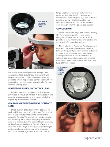
POSTERIOR FUNDUS CONTACT LENS
This is a modified Koeppe lens. The image
produced is virtual and erect. It is situated in the
anterior vitreous cavity and is most helpful in
exploring the posterior fundus.
GOLDMANN THREE-MIRROR CONTACT
LENS
Three mirrors are placed in the cone, each
with a different angle of inclination. The central
part of the contact lens allows a view of the
posterior fundus. The peripheral fundus can be
examined by each mirror, bringing a different
area into the fundus. The basis of biomicroscopy
of the vitreous body is the Tyndall effect. It is
maximal with a high intensity of projected light
and a good contrast between the observed
structure and background. It also includes a
119 | OPTOMETRY