
Page 122 - The Indian Optician Digital Edition November-December 2021
P. 122
EXAMINATION OF THE
FUNDUS BY
FOCAL
ILLUMINATION
he ordinary slit-lamp cannot be used to explore
the eye further back than the anterior parts of the
Tvitreous. This is due to the beam of light being
ordinarily brought to a focus in this region. However,
there are instances where the beam is made more
divergent by eliminating the refractive influence of the
corneal curvature. This can be done by using a contact
lens with a flat anterior face or by interposing a -55 d
lens in front of the cornea. In that case, the posterior
part of the vitreous and the central area of the fundus is
Dr Vineet Shrivastava examined with the help of a binocular microscope and
B.E.M.S., D.O., (FOOREC) a focused beam of light.
FCLC (Gold Medalist)
Consultant Optometrist & Educator This kind of examination requires full mydriasis. By
Banswara, Rajasthan
this method, fine changes in the posterior part
of the vitreous, the retina, and the optic disc
can be readily studied. The areas of oedema
are clearly outlined in the optical section,
thus making it an effective process. Problems
in diagnosis, such as the difference between
a cyst and a hole at the macula, are properly
demonstrated as well. Three types of lenses are
available for a biomicroscopic examination of the
vitreous and fundus - Hruby lens, Posterior Fundus
contact lens and Goldmann Three-Mirror contact lens.
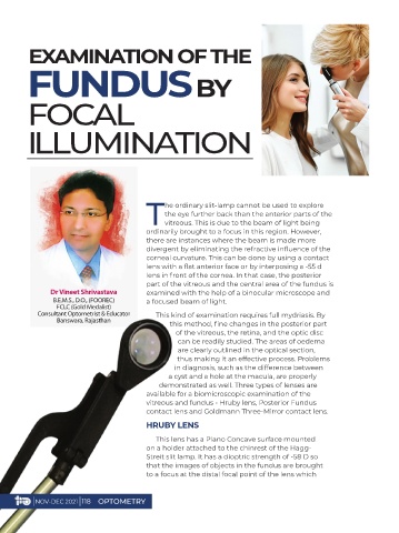
HRUBY LENS
This lens has a Plano Concave surface mounted
on a holder attached to the chinrest of the Hagg-
Streit slit lamp. It has a dioptric strength of -58 D so
that the images of objects in the fundus are brought
to a focus at the distal focal point of the lens which
| NOV-DEC 2021 | 118 OPTOMETRY