
Page 171 - July-August 2020
P. 171
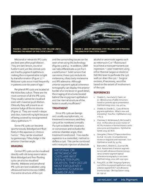
FIGURE 3.  UBM OF PERIPHERAL CYST. YELLOW LINE IS   FIGURE 4.  UBM OF MIDZONAL CYST. YELLOW LINE IS TRACING
            TRACING THE HEIGHT OF THE CYSTIC CAVITY            THE LENGTH OF THE CYSTIC CAVITY


             Midzonal or retroiridic IPE cysts   and the surrounding tissues can be   alcohol or antimitotic agents such
          are best seen after pupil dilation.    seen when using this technology   as mitomycin-C or  -fluorouracil
                                                                                               5
          They are dark brown, round or     (Figures 3 and 4).  In addition, UBM   to achieve a more permanent cyst
          fusiform and just like central cysts   can help differentiate a cyst from   regression.   Laser treatment using
                                                                                        4
          are lined by pigment epithelium   a solid tumor.   Solid tumors that   either thermal (argon or diode) or
                                                        5
          making them impenetrable to light   can mimic these cysts include iris   Nd:YAG laser to perforate the cyst
          by transillumination (Figure 1).    melanoma, ciliary body melanoma   wall can drain the cyst.   Surgical
                                                                                                  4
                                    2,4
          Midzonal cysts occur most frequently  and IPE adenoma. Although      excision, if necessary, would be
          in patients over 60 years of age. 1  anterior segment optical coherence   based on the extent of involvement
                                            tomography can display the anterior   of the cyst.
             Peripheral IPE cysts are located at   border of an iris lesion in good detail,
          the iridociliary sulcus. These are the   the imaging of structures located   REFERENCES
          most common of all the IPE cysts.    behind the iris pigment epithelium   1.  Shields CL, Kancherla S, Patel J, et
          They usually cannot be visualised   and the internal structure of the   al.  Clinical survey of 3680 iris tumors
          even with maximal pupil dilation.   lesion is usually insufficient.     based on patients age at presentation.
                                                                    6
          Clinically they will present as an                                      Ophthalmology 2012; 119: 407-14.
          anterior bulge of the iris stroma   TREATMENT                        2.  Shields JA, Shields CL.  Cysts of the iris
          (Figure 2).  They are round in shape                                    pigment epithelium.  What is new and
          and clear, transmitting light because   Since IPE cysts are benign      interesting?  Asia-Pac J Ophthalmol 2017;
          of being covered by nonpigmented   and usually asymptomatic, no         1: 64-9.
          ciliary epithelium. 2,4           treatment is necessary and they    3.  Chamney S, McGimpsey S, McConnell V,
                                            can just be monitored annually.       et al.  Iris flocculi as an ocular marker of
             Some IPE cysts can become      If a cyst occludes the visual axis    ACTA2 mutation in familial thoracic aortic
          spontaneously dislodged and float   or compresses and occludes the      aneurysms and dissections.  Ophthalmic
          freely in the aqueous or vitreous   anterior chamber angle, then        Genet 2015; 36: 86-8.
          humor.   They can be visualised   treatment is indicated.  Fine-needle   4.  Georgalas I, Petrou P, Papaconstantinou
                2
          by gonioscopy if they lodge in the   aspiration is a controlled, method to   D, et al.  Iris cysts:  A comprehensive
                                                                                  review on diagnosis and treatment.  Surv
          anterior chamber angle.           deflate a cyst.  This can be combined   Ophthalmol 2018; 63: 347-64.
                                            with intracystic injection of absolute
          IMAGING                                                              5.  Bianciotto C, Shields CL, Guzman JM,
                                                                                  et al.  Assessment of anterior segment
             Central IPE cysts can be visualised   MIDZONAL OR                    tumors with ultrasound biomicroscopy
          directly with slit-lamp examination.    RETROIRIDIC IPE                 versus anterior segment optical
          Most dislodged and free-floating    CYSTS ARE BEST                      coherence tomography in 200 cases.
          cysts can also be visualised        SEEN AFTER PUPIL                    Ophthalmology 2011; 118: 1297-1302.
          directly.  Midzone and peripheral   DILATION.  THEY OCCUR            6.  Pong JCF, Lai JSM.  Imaging of primary
                                                                                  cyst of the iris pigment epithelium using
          IPE cysts are best evaluated using   MOST FREQUENTLY                    anterior segment OCT and ultrasonic
          ultrasound biomicroscopy (UBM).     IN PATIENTS OVER 60                 biomicroscopy.  Clin Exp Optom 2009;
          The entire structure of the cyst    YEARS OF AGE                        92: 139-41.

                                                                                  JULY-AUG 2020  |  167  Iris Pigment