
Page 170 - July-August 2020
P. 170
IRIS PIGMENT
EPITHELIAL CYSTS
Primary iris cysts arise from the iris pigment epithelium
(IPE) or the iris stroma. The more posteriorly located IPE
cysts are subdivided according to their location: central
(pupillary margin), midzonal (retroiridic), peripheral
(iridociliary) and dislodged (free-floating). The most
common type of iris cysts are IPE cysts and these will be
reviewed in this article.
Leonid Skorin ANATOMY
Jr., O.D., D.O., M.S.
The iris consists of two layers: the stroma anteriorly and the pigment
Mayo Clinic Health System epithelium posteriorly. Primary IPE cysts are composed of neuroepithelium.
Consultant, Department of Surgery
Community Division of Ophthalmology Up to 85% of all primary iris cysts are IPE cysts. 1
Albert Lea, Minnesota
Primary cysts of the IPE are subdivided anatomically into four types. The
central or pupillary margin IPE cysts are visible without dilation. They are
dark brown in colour and teardrop in shape. They most often can be found
2
in both eyes. Iris flocculi are a unique form of central IPE cysts and are
associated with increased risk of dissecting thoracic aortic aneurysms. 3
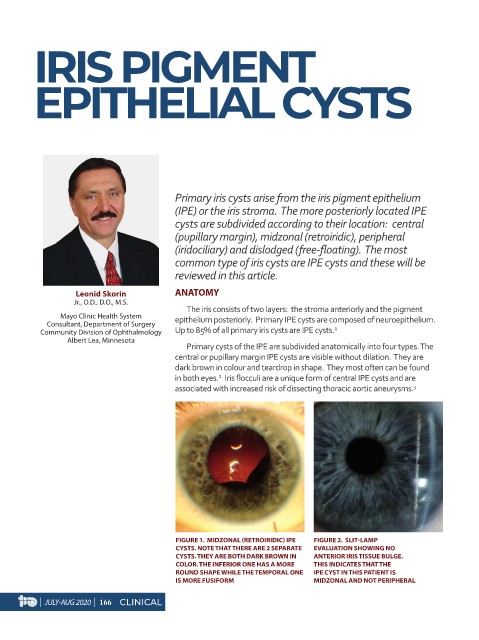
FIGURE 1. MIDZONAL (RETROIRIDIC) IPE FIGURE 2. SLIT-LAMP
CYSTS. NOTE THAT THERE ARE 2 SEPARATE EVALUATION SHOWING NO
CYSTS. THEY ARE BOTH DARK BROWN IN ANTERIOR IRIS TISSUE BULGE.
COLOR. THE INFERIOR ONE HAS A MORE THIS INDICATES THAT THE
ROUND SHAPE WHILE THE TEMPORAL ONE IPE CYST IN THIS PATIENT IS
IS MORE FUSIFORM MIDZONAL AND NOT PERIPHERAL
| JULY-AUG 2020 | 166 CLINICAL