
Page 101 - The Indian Optician Digital Edition November-December 2021
P. 101
with laser refractive surgery in myopic
and hyperopic patients, as well as possible
subsequent cataract interventions. We are
suggesting something totally new that is also
not incompatible with any other ocular therapy.”
This inlay, unlike others that do exist, would
not prevent subsequent study of the retina
or macula and even surgical interventions,
according to Dr Salvador García-Delpech from
the Aiken Foundation.
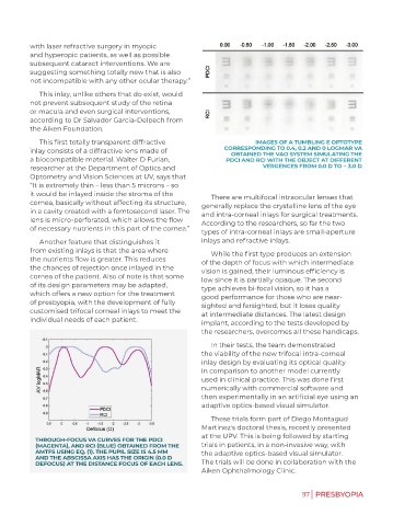
This first totally transparent diffractive IMAGES OF A TUMBLING E OPTOTYPE
inlay consists of a diffractive lens made of CORRESPONDING TO 0.4, 0.2 AND 0 LOGMAR VA
OBTAINED THE VAO SYSTEM SIMULATING THE
a biocompatible material. Walter D Furlan, PDCI AND RCI WITH THE OBJECT AT DIFFERENT
researcher at the Department of Optics and VERGENCES FROM 0.0 D TO − 3.0 D
Optometry and Vision Sciences at UV, says that
“It is extremely thin – less than 5 microns – so
it would be inlayed inside the stroma of the There are multifocal intraocular lenses that
cornea, basically without affecting its structure, generally replace the crystalline lens of the eye
in a cavity created with a femtosecond laser. The and intra-corneal inlays for surgical treatments.
lens is micro-perforated, which allows the flow According to the researchers, so far the two
of necessary nutrients in this part of the cornea.” types of intra-corneal inlays are small-aperture
Another feature that distinguishes it inlays and refractive inlays.
from existing inlays is that the area where While the first type produces an extension
the nutrients flow is greater. This reduces of the depth of focus with which intermediate
the chances of rejection once inlayed in the vision is gained, their luminous efficiency is
cornea of the patient. Also of note is that some low since it is partially opaque. The second
of its design parameters may be adapted, type achieves bi-focal vision, so it has a
which offers a new option for the treatment good performance for those who are near-
of presbyopia, with the development of fully sighted and farsighted, but it loses quality
customised trifocal corneal inlays to meet the at intermediate distances. The latest design
individual needs of each patient. implant, according to the tests developed by
the researchers, overcomes all these handicaps.
In their tests, the team demonstrated
the viability of the new trifocal intra-corneal
inlay design by evaluating its optical quality
in comparison to another model currently
used in clinical practice. This was done first
numerically with commercial software and
then experimentally in an artificial eye using an
adaptive optics-based visual simulator.
These trials form part of Diego Montagud
Martínez's doctoral thesis, recently presented
at the UPV. This is being followed by starting
THROUGH-FOCUS VA CURVES FOR THE PDCI
(MAGENTA), AND RCI (BLUE) OBTAINED FROM THE trials in patients, in a non-invasive way, with
AMTFS USING EQ. (1). THE PUPIL SIZE IS 4.5 MM the adaptive optics-based visual simulator.
AND THE ABSCISSA AXIS HAS THE ORIGIN (0.0 D
DEFOCUS) AT THE DISTANCE FOCUS OF EACH LENS. The trials will be done in collaboration with the
Aiken Ophthalmology Clinic.
97 | PRESBYOPIA