
Page 92 - The Indian Digital Edition September-October 2020
P. 92
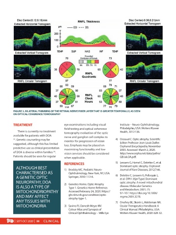
FIGURE 3. BILATERAL THINNING OF THE RETINAL NERVE FIBER LAYER THAT IS GREATER TEMPORALLY, AS SEEN
          ON OPTICAL COHERENCE TOMOGRAPHY


          TREATMENT                         eye examinations including visual     Institute -- Neuro-Ophthalmology.
                                            field testing and optical coherence   Philadelphia, USA: Wolters Kluwer
             There is currently no treatment   tomography evaluation of the optic   Health, 2012:136.
          available for patients with DOA   nerve and ganglion cell complex to
          (3) . Genetic counseling may be   monitor for progression of vision   4)  Orssaud C. Optic atrophy. Scientific
          suggested, although this has limited   loss. Emphasis may be placed on   Editor: Professor Jean-Louis Dufier.
          predictive use as clinical presentation   maximising functionality and low   Orphanet Encyclopedia; November
                                                                                  2003. Accessed  March 2, 2020.
          of DOA is diverse within families  .   vision services should be considered   http://www.orpha.net/data/patho/
                                     (6)
          Patients should be seen for regular   when applicable.                  GB/uk.OA.pdf.
                                            REFERENCES                         5)  Lenaers G, Hamel C, Delettre C, et al.
            ALTHOUGH BEST                   1)  Brodsky MC. Pediatric Neuro-      Dominant optic atrophy. Orphanet
                                                                                  Journal of Rare Diseases; 2012;7:46.
            CHARACTERISED AS                   Ophthalmology. New York, NY, USA:
            A GENETIC OPTIC                    Springer, 2010: 172-6.          6)  Delettre C, Lenaers G, Pelloquin L,
                                                                                  et al. OPA1 (Kjer Type) Dominant
            NEUROPATHY, DOA                 2)  Genetics Home. Optic Atrophy      optic atrophy: A novel mitochondrial
            IS ALSO A TYPE OF                  Type 1. Genetics Home Reference.   disease. Molecular Genetics
            MITOCHONDRIOPATHY                  Accessed February 24, 2020. https://  and Metabolism; 2001; 75:
                                                                                  97–107. https://doi.org/10.1006/
            AND MAY AFFECT                     ghr.nlm.nih.gov/condition/optic-   mgme.2001.3278.
                                               atrophy-type-1.
            ANY TISSUES WITH                                                   7)  Onofrey BE, Skorin L, Holdeman NR.
            MITOCHONDRIA                    3)  Savino PJ, Danesh-Meyer HV.       Ocular Therapeutics Handbook: A
                                               Colour Atlas and Synopsis of       Clinical Manual. Philadelphia, USA:
                                               Clinical Ophthalmology -- Wills Eye   Wolters Kluwer Health, 2020: 628-32.

           |  SEPT-OCT 2020  |  88  CLINICAL