
Page 142 - July-August 2020
P. 142
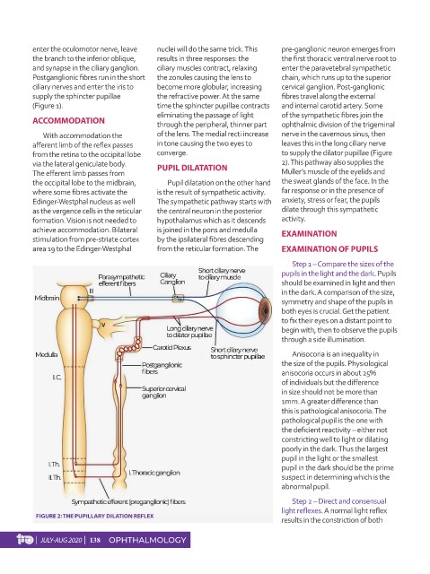
enter the oculomotor nerve, leave   nuclei will do the same trick. This   pre-ganglionic neuron emerges from
          the branch to the inferior oblique,   results in three responses: the   the first thoracic ventral nerve root to
          and synapse in the ciliary ganglion.   ciliary muscles contract, relaxing   enter the paravetebral sympathetic
          Postganglionic fibres run in the short   the zonules causing the lens to   chain, which runs up to the superior
          ciliary nerves and enter the iris to   become more globular, increasing   cervical ganglion. Post-ganglionic
          supply the sphincter pupillae     the refractive power. At the same   fibres travel along the external
          (Figure 1).                       time the sphincter pupillae contracts   and internal carotid artery. Some
                                            eliminating the passage of light   of the sympathetic fibres join the
          ACCOMMODATION                     through the peripheral, thinner part   ophthalmic division of the trigeminal
             With accommodation the         of the lens. The medial recti increase   nerve in the cavernous sinus, then
          afferent limb of the reflex passes   in tone causing the two eyes to   leaves this in the long ciliary nerve
          from the retina to the occipital lobe   converge.                    to supply the dilator pupillae (Figure
          via the lateral geniculate body.   PUPIL DILATATION                  2). This pathway also supplies the
          The efferent limb passes from                                        Muller’s muscle of the eyelids and
          the occipital lobe to the midbrain,   Pupil dilatation on the other hand   the sweat glands of the face. In the
          where some fibres activate the    is the result of sympathetic activity.   far response or in the presence of
          Edinger-Westphal nucleus as well   The sympathetic pathway starts with   anxiety, stress or fear, the pupils
          as the vergence cells in the reticular   the central neuron in the posterior   dilate through this sympathetic
          formation. Vision is not needed to   hypothalamus which as it descends   activity.
          achieve accommodation. Bilateral   is joined in the pons and medulla   EXAMINATION
          stimulation from pre-striate cortex   by the ipsilateral fibres descending
          area 19 to the Edinger-Westphal   from the reticular formation. The   EXAMINATION OF PUPILS

                                                                                  Step 1 – Compare the sizes of the
                                                                               pupils in the light and the dark. Pupils
                                                                               should be examined in light and then
                                                                               in the dark. A comparison of the size,
                                                                               symmetry and shape of the pupils in
                                                                               both eyes is crucial. Get the patient
                                                                               to fix their eyes on a distant point to
                                                                               begin with, then to observe the pupils
                                                                               through a side illumination.
                                                                                  Anisocoria is an inequality in
                                                                               the size of the pupils. Physiological
                                                                               anisocoria occurs in about 25%
                                                                               of individuals but the difference
                                                                               in size should not be more than
                                                                               1mm. A greater difference than
                                                                               this is pathological anisocoria. The
                                                                               pathological pupil is the one with
                                                                               the deficient reactivity – either not
                                                                               constricting well to light or dilating
                                                                               poorly in the dark. Thus the largest
                                                                               pupil in the light or the smallest
                                                                               pupil in the dark should be the prime
                                                                               suspect in determining which is the
                                                                               abnormal pupil.

                                                                                  Step 2 – Direct and consensual
                                                                               light reflexes. A normal light reflex
           FIGURE 2: THE PUPILLARY DILATION REFLEX
                                                                               results in the constriction of both

           |  JULY-AUG 2020  |  138  OPHTHALMOLOGY