
Page 146 - July-August 2020
P. 146
This could be due to an intraocular SINGLE SMALL PUPIL
tumour, formation of anterior PUPIL SIZE IS
synechiae or posterior synechiae A RESULT OF HORNER’S SYNDROME
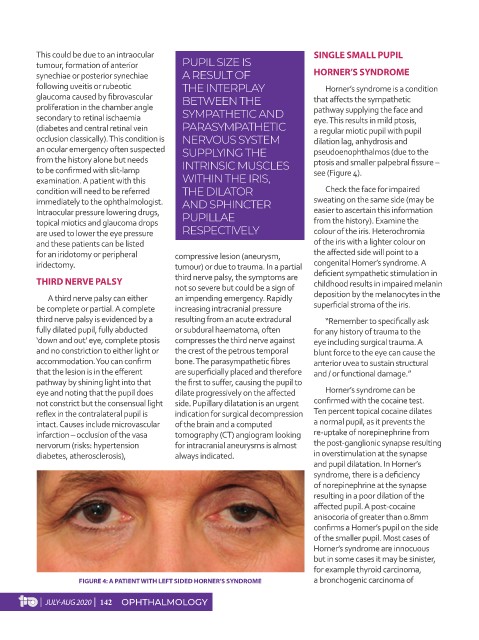
following uveitis or rubeotic THE INTERPLAY Horner’s syndrome is a condition
glaucoma caused by fibrovascular BETWEEN THE that affects the sympathetic
proliferation in the chamber angle pathway supplying the face and
secondary to retinal ischaemia SYMPATHETIC AND eye. This results in mild ptosis,
(diabetes and central retinal vein PARASYMPATHETIC a regular miotic pupil with pupil
occlusion classically). This condition is NERVOUS SYSTEM dilation lag, anhydrosis and
an ocular emergency often suspected SUPPLYING THE pseudoenophthalmos (due to the
from the history alone but needs ptosis and smaller palpebral fissure –
to be confirmed with slit-lamp INTRINSIC MUSCLES see (Figure 4).
examination. A patient with this WITHIN THE IRIS,
condition will need to be referred THE DILATOR Check the face for impaired
immediately to the ophthalmologist. AND SPHINCTER sweating on the same side (may be
Intraocular pressure lowering drugs, PUPILLAE easier to ascertain this information
topical miotics and glaucoma drops from the history). Examine the
are used to lower the eye pressure RESPECTIVELY colour of the iris. Heterochromia
and these patients can be listed of the iris with a lighter colour on
for an iridotomy or peripheral compressive lesion (aneurysm, the affected side will point to a
iridectomy. tumour) or due to trauma. In a partial congenital Horner’s syndrome. A
THIRD NERVE PALSY third nerve palsy, the symptoms are deficient sympathetic stimulation in
childhood results in impaired melanin
not so severe but could be a sign of
A third nerve palsy can either an impending emergency. Rapidly deposition by the melanocytes in the
be complete or partial. A complete increasing intracranial pressure superficial stroma of the iris.
third nerve palsy is evidenced by a resulting from an acute extradural “Remember to specifically ask
fully dilated pupil, fully abducted or subdural haematoma, often for any history of trauma to the
‘down and out’ eye, complete ptosis compresses the third nerve against eye including surgical trauma. A
and no constriction to either light or the crest of the petrous temporal blunt force to the eye can cause the
accommodation. You can confirm bone. The parasympathetic fibres anterior uvea to sustain structural
that the lesion is in the efferent are superficially placed and therefore and / or functional damage.”
pathway by shining light into that the first to suffer, causing the pupil to
eye and noting that the pupil does dilate progressively on the affected Horner’s syndrome can be
not constrict but the consensual light side. Pupillary dilatation is an urgent confirmed with the cocaine test.
reflex in the contralateral pupil is indication for surgical decompression Ten percent topical cocaine dilates
intact. Causes include microvascular of the brain and a computed a normal pupil, as it prevents the
infarction – occlusion of the vasa tomography (CT) angiogram looking re-uptake of norepinephrine from
nervorum (risks: hypertension for intracranial aneurysms is almost the post-ganglionic synapse resulting
diabetes, atherosclerosis), always indicated. in overstimulation at the synapse
and pupil dilatation. In Horner’s
syndrome, there is a deficiency
of norepinephrine at the synapse
resulting in a poor dilation of the
affected pupil. A post-cocaine
anisocoria of greater than 0.8mm
confirms a Horner’s pupil on the side
of the smaller pupil. Most cases of
Horner’s syndrome are innocuous
but in some cases it may be sinister,
for example thyroid carcinoma,
FIGURE 4: A PATIENT WITH LEFT SIDED HORNER’S SYNDROME a bronchogenic carcinoma of
| JULY-AUG 2020 | 142 OPHTHALMOLOGY