
Page 140 - July-August 2020
P. 140
THE ASSESSMENT OF
PUPILS AND PUPILLARY
REACTIONS
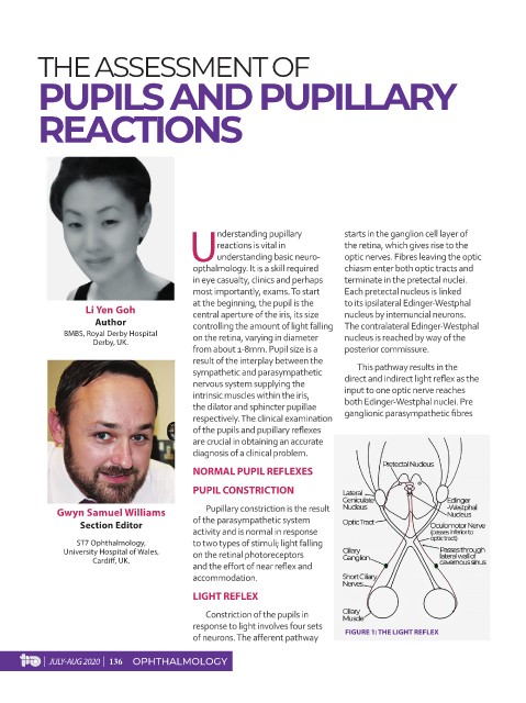
nderstanding pupillary starts in the ganglion cell layer of
reactions is vital in the retina, which gives rise to the
U understanding basic neuro- optic nerves. Fibres leaving the optic
opthalmology. It is a skill required chiasm enter both optic tracts and
in eye casualty, clinics and perhaps terminate in the pretectal nuclei.
most importantly, exams. To start Each pretectal nucleus is linked
at the beginning, the pupil is the to its ipsilateral Edinger-Westphal
Li Yen Goh central aperture of the iris, its size nucleus by internuncial neurons.
Author controlling the amount of light falling The contralateral Edinger-Westphal
BMBS, Royal Derby Hospital on the retina, varying in diameter nucleus is reached by way of the
Derby, UK.
from about 1-8mm. Pupil size is a posterior commissure.
result of the interplay between the
sympathetic and parasympathetic This pathway results in the
nervous system supplying the direct and indirect light reflex as the
intrinsic muscles within the iris, input to one optic nerve reaches
the dilator and sphincter pupillae both Edinger-Westphal nuclei. Pre
respectively. The clinical examination ganglionic parasympathetic fibres
of the pupils and pupillary reflexes
are crucial in obtaining an accurate
diagnosis of a clinical problem.
NORMAL PUPIL REFLEXES
PUPIL CONSTRICTION
Gwyn Samuel Williams Pupillary constriction is the result
Section Editor of the parasympathetic system
activity and is normal in response
ST7 Ophthalmology, to two types of stimuli; light falling
University Hospital of Wales, on the retinal photoreceptors
Cardiff, UK.
and the effort of near reflex and
accommodation.
LIGHT REFLEX
Constriction of the pupils in
response to light involves four sets
of neurons. The afferent pathway FIGURE 1: THE LIGHT REFLEX
| JULY-AUG 2020 | 136 OPHTHALMOLOGY