
Page 168 - The Indian Optician Digital Edition Jan-Feb 2020
P. 168
MACULAR TELANGIECTASIA juxtafoveolar retinal telangiectasis.
TYPE 3 Update of classification and follow-
up study. Ophthalmology 1993;
Macular telangiectasia type 100:1536-46.
3 (MacTel type 3), also known as 2. Panos G, et al. The diagnosis
occlusive telangiectasia is a rare and management of macular
form of macular telangiectasia and telangiectasia. Ophthal Surg Laser
is least understood. It is caused Imag Retina. 2019;50:139-144.
by capillary nonperfusion and 3. Yannuzzi L, et al. Idiop Mac
can have microaneurysms. MacTel Telang. Arch Ophthalmol.
2006;124:450-460.
type 3 has been associated with
3
vasculopathy in the brain. MRI 4. Osaka, et al. Clinical features
imaging with angiography is of treated and untreated type 1
idiopathic macular telangiectasia
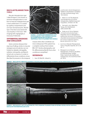
necessary if detected. FIGURE 7 - FLUORESCEIN ANGIOGRAPHY: LEFT without the occurence of secondary
EYE WITH MACTEL TYPE 2 SHOWING POOLING
OF FLUORESCEIN WITHIN THE MACULA choroidal neovascularization followed
DIFFERENTIAL DIAGNOSIS TEMPORALLY DUE TO TELANGIECTATIC VESSELS for 2 years in Japanese patients.
AND CONCLUSION Retina: 2018; 38: S114–S122.
macula while other conditions are
Some common diseases that more widespread. If the patient has 5. Issa P, et al. Macular Telangiectasia
may have findings similar to macular a complete workup that includes Type 2. Prog Retin Eye Res. 2013; 34:
telangiectasia include but are not BIO, OCT, fundus photography, and 49–77.
limited to: diabetic retinopathy, FA, differentiating MacTel from other 6. Mozayan E, et al. Macular
radiation retinopathy, and branch conditions is simplified. 5 telangiectasia. American Academy
retinal vein occlusions. In order to of Ophthalmology. EyeWiki.
https://eyewiki.aao.org/Macular_
differentiate these conditions, know REFERENCES telangiectasia. Accessed November
that MacTel presents in the temporal 1. Gass JD, Blodi BA. Idiopathic 20 2019.
th
FIGURE 5 - MACULAR OCT: RIGHT EYE SHOWING A PSEUDOLAMELLAR HOLE DUE TO ATROPHY IN THE LATER STAGES OF MACTEL TYPE 2
FIGURE 6 - MACULAR OCT: LEFT EYE WITH MACTEL TYPE 2 SHOWING TELANGIECTASIA OF RETINAL VESSELS IN THE TEMPORAL
MACULA OF THE INNER NUCLEAR LAYER
| JAN-FEB 2020 |164 CLINICAL
January-February 2020 SK.indd 77 02/15/2020 16:09