
Page 166 - The Indian Optician Digital Edition Jan-Feb 2020
P. 166
MACULAR
TELANGIECTASIAS:
SUBTLE, YET SIGNIFICANT
INTRODUCTION
First described by Gass and Blodi, macular telangiectasias
(MacTel) are part of a group of conditions referred to as idiopathic
juxtafoveolar retinal telangiectasias. These telangiectasias are
classified into three groups. This article reviews and identifies how
each of these groups are managed. 1
MACULAR TELANGIECTASIA to detect these retinal findings,
TYPE 1 full fundus examination is needed.
Binocular indirect ophthalmoscopy
Macular telangiectasia type (BIO), fundus photography, optical
1 (MacTel type 1) is an idiopathic coherence tomography (OCT), and
Taylor Lauermann juxtafoveolar telangiectasia,
BS fluorescein angiography (FA) should
commonly referred to as aneurysmal be performed in these patients.
4 year Optometry Student telangiectasia. This condition is
th
Pacific University College of Optometry Current research is looking at
Forest Grove, Oregon considered a variant of Coats disease anti-vascular endothelial growth
and is unilateral, congenital, and factor (anti-VEGF) injections and
normally presents in middle-aged laser photocoagulation as possible
men. MacTel type 1 is located treatment for these patients. 2,4
temporal to the macula with
aneurysms of the capillaries,
veins, and arteries. These
vessels are leaky and
therefore can cause edema
and hard exudates to
appear within the macula.
2,3
If the edema is significant
within the macula it can
lead to vision loss. In order
Leonid Skorin
Jr., O.D., D.O., M.S.
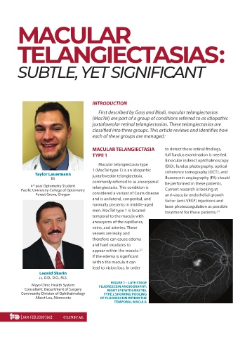
FIGURE 1 - LATE STAGE
Mayo Clinic Health System FLUORESCEIN ANGIOGRAPHY:
Consultant, Department of Surgery RIGHT EYE WITH MACTEL
Community Division of Ophthalmology TYPE 2 SHOWING POOLING
Albert Lea, Minnesota OF FLUORESCEIN WITHIN THE
TEMPORAL MACULA
| JAN-FEB 2020 |162 CLINICAL
January-February 2020 SK.indd 75 02/15/2020 16:08